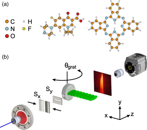
Figure 1
(a) The experiments are performed with the antibiotic ciprofloxacin (left) and the organic dye phthalocyanine (right). (b) A thermal beam of molecules is produced by microevaporation and collimated vertically (Sx) and horizontally (Sy). After 1.5 m of free flight the molecules are diffracted at a thick laser grating created by retroreflecting a 532 nm laser at a highly reflective mirror. The angle of the mirror with respect to the molecular beam θgrat can be controlled with μ rad precision. The molecular diffraction pattern is recorded after further 0.57 m of free flight by laser-induced fluorescence microscopy.
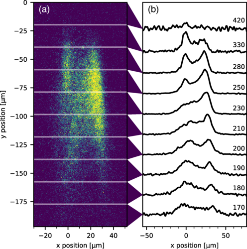
Figure 3
Bragg diffraction pattern of the organic dye molecule phthalocyanine at an incidence angle θgrat=5(5) μrad. Panel (a) shows the false color diffraction image. Panel (b) shows the averages of 20 μm high stripes, smoothed with median and Savitzky–Golay [38] filters, and annotated with their corresponding velocities in m/s. The velocities are determined by comparison with a diffraction pattern produced by a material grating [21]. The laser grating waist for this measurement is wy=57(3) μm and the collimation slit width is 11.5 μm.
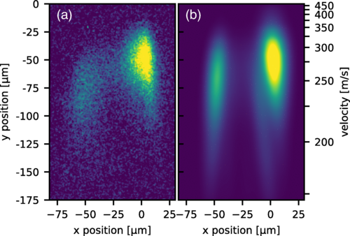
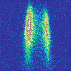
Figure 2
False color image of the experimental (a) and simulated (b) Bragg diffraction pattern of the antibiotic ciprofloxacin. The laser grating waists are wz=7.04(5) mm, wy=55(5) μm, and the collimation slit is set to 14 μm.
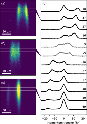
Figure 4
Angular dependence of Bragg diffraction of the dye molecule phthalocyanine. Panels (a)–(c) show diffraction images for the incidence angles 48 (a), −5 (b) and −69 μrad (c). The images are 197 by 197 μm and the scale bars are 50 μm long. Panel (d) shows the integrated intensity profiles for the incidence angle varying from −69 to 48 μrad in steps of 10 μrad. The profiles are averages of 16 μm high stripes of the diffraction images corresponding to a velocity range of 234 to 255 m/s. The curves are horizontally aligned to center the undiffracted beam (which is the right peak for negative incidence and the left peak for positive incidence). For ±5 μrad we observe diffraction to both sides of the initial beam and hence align the traces with respect to their center of gravity. The laser grating waist for this measurement is wy=65(5) μm and the collimation slit width is 14.8 μm.
Figure 1
(a) The experiments are performed with the antibiotic ciprofloxacin (left) and the organic dye phthalocyanine (right). (b) A thermal beam of molecules is produced by microevaporation and collimated vertically (Sx) and horizontally (Sy). After 1.5 m of free flight the molecules are diffracted at a thick laser grating created by retroreflecting a 532 nm laser at a highly reflective mirror. The angle of the mirror with respect to the molecular beam θgrat can be controlled with μ rad precision. The molecular diffraction pattern is recorded after further 0.57 m of free flight by laser-induced fluorescence microscopy.
Figure 2
False color image of the experimental (a) and simulated (b) Bragg diffraction pattern of the antibiotic ciprofloxacin. The laser grating waists are wz=7.04(5) mm, wy=55(5) μm, and the collimation slit is set to 14 μm.
Figure 3
Bragg diffraction pattern of the organic dye molecule phthalocyanine at an incidence angle θgrat=5(5) μrad. Panel (a) shows the false color diffraction image. Panel (b) shows the averages of 20 μm high stripes, smoothed with median and Savitzky–Golay [38] filters, and annotated with their corresponding velocities in m/s. The velocities are determined by comparison with a diffraction pattern produced by a material grating [21]. The laser grating waist for this measurement is wy=57(3) μm and the collimation slit width is 11.5 μm.
Figure 4
Angular dependence of Bragg diffraction of the dye molecule phthalocyanine. Panels (a)–(c) show diffraction images for the incidence angles 48 (a), −5 (b) and −69 μrad (c). The images are 197 by 197 μm and the scale bars are 50 μm long. Panel (d) shows the integrated intensity profiles for the incidence angle varying from −69 to 48 μrad in steps of 10 μrad. The profiles are averages of 16 μm high stripes of the diffraction images corresponding to a velocity range of 234 to 255 m/s. The curves are horizontally aligned to center the undiffracted beam (which is the right peak for negative incidence and the left peak for positive incidence). For ±5 μrad we observe diffraction to both sides of the initial beam and hence align the traces with respect to their center of gravity. The laser grating waist for this measurement is wy=65(5) μm and the collimation slit width is 14.8 μm.